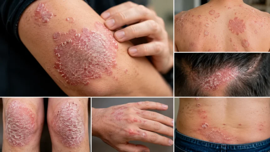
Pictures of Psoriasis: Types, Symptoms and Skin Changes
Psoriasis can look very different from one person to another, which is why people often search for pictures of psoriasis when an unfamiliar rash, patch, or scaling develops. Some people have thick plaques covered with silvery scale, while others develop small drop-shaped spots, smooth patches in skin folds, pustules, scalp scaling, or changes in their fingernails and toenails. Skin tone also affects how inflammation appears, so psoriasis is not always bright red. On darker skin, affected areas may appear purple, violet, gray, brown, or darker than the surrounding skin. Knowing these variations can make psoriasis easier to recognize, but photographs should be used as a guide rather than a substitute for medical diagnosis.
Psoriasis is a chronic immune-mediated condition in which skin cells are produced much faster than usual, causing them to accumulate on the surface. The National Institute of Arthritis and Musculoskeletal and Skin Diseases explains that psoriasis commonly causes inflamed, scaly patches and may go through periods of flares followed by quieter periods. The condition is not contagious, so it cannot spread through touching someone who has psoriasis. Different types can occur in different areas of the body, and some people develop more than one form during their lifetime. Understanding the characteristic psoriasis symptoms and skin changes can help you know when a rash deserves professional evaluation.
What Does Psoriasis Look Like?
The classic picture of psoriasis shows a clearly defined patch of thickened skin with scale on its surface. In plaque psoriasis, the most common form, these areas may be raised and covered with white, silver, or gray scales that can flake away. The American Academy of Dermatology reports that plaque psoriasis occurs in approximately 80% to 90% of people with psoriasis. Common locations include the knees, elbows, scalp, and lower back, although plaques can develop almost anywhere. Patches can be small and isolated or join together into larger areas of affected skin. Itching is common, and some plaques become sore, cracked, or painful when the inflammation is more severe.
The color of psoriasis varies significantly with natural skin tone, which is important when comparing your skin with online psoriasis pictures. On lighter skin, inflammation is often described as pink or red, frequently beneath a white or silvery scale. On medium or darker skin, psoriasis may look purple, violet, dark brown, gray, or deeper than the surrounding skin rather than obviously red. The scale can still appear pale, grayish, or silvery, making the borders easier to notice. After a flare improves, darker or lighter discoloration may remain in the affected area for some time. Because many traditional medical images historically emphasized lighter skin tones, people with darker skin may not always recognize their symptoms in commonly displayed examples.
Texture is often just as useful as color when identifying possible psoriasis. A plaque may feel noticeably thicker than nearby skin and may have a dry, rough, layered surface. Repeated scratching or rubbing can loosen scale and sometimes cause tiny spots of bleeding when the underlying skin is irritated. Psoriasis may also burn, sting, itch, or feel tight, particularly if the skin becomes very dry. In areas where the skin naturally folds, however, the condition can appear smooth and shiny rather than thick and scaly. This difference is one reason looking at photographs of only plaque psoriasis can be misleading. The appearance depends greatly on the psoriasis type and the body area involved.
Another clue is the pattern in which lesions appear. Plaque psoriasis often affects both sides of the body in a roughly symmetrical pattern, such as both elbows or both knees, although symmetry is not required for diagnosis. Guttate psoriasis may instead create dozens of small spots scattered across the torso and limbs. Inverse psoriasis typically develops within folds, while palmoplantar forms affect the palms or soles. Scalp psoriasis can stay within the hairline or extend beyond it onto the forehead, ears, or neck. Nail psoriasis may occur even when skin plaques are limited or absent. Paying attention to location and pattern can therefore be more informative than focusing on color alone.
Pictures can help narrow possibilities, but several common conditions resemble psoriasis. Eczema may produce inflamed, itchy, scaly patches, while fungal infections can create sharply bordered rashes that are sometimes mistaken for psoriasis. Seborrheic dermatitis can resemble scalp psoriasis, and irritation or allergic contact dermatitis may create patches in many of the same locations. Healthcare professionals usually diagnose psoriasis by examining the skin, scalp, and nails while considering personal and family history. Occasionally, a small skin sample may be examined when another condition needs to be excluded. For that reason, a photograph can provide a useful comparison but cannot confirm whether a particular rash is psoriasis.
Pictures of Plaque Psoriasis and Its Common Symptoms
Plaque psoriasis is the form most people picture when they hear the word psoriasis. It usually creates raised, clearly defined areas of inflamed skin covered by visible scale. These plaques can range from small coin-sized patches to large connected areas that cover substantial portions of the body. The scale often appears silvery white on lighter skin and may look grayish on darker skin. Common locations include the elbows, knees, scalp, trunk, and lower back, although any skin surface can potentially be affected. NIAMS describes plaque psoriasis as the most common form and notes that lesions often develop in a symmetrical pattern.
Early plaque psoriasis may look less dramatic than photographs of established disease. A person might initially notice a small dry patch that repeatedly flakes and returns despite ordinary moisturizers. As inflammation increases, the border may become more distinct and the skin beneath the scale can become thicker. Multiple small plaques may eventually enlarge or merge into one broader patch. Some lesions remain relatively stable, while others expand during a flare and gradually flatten when treatment begins working. Psoriasis commonly follows a relapsing pattern, which means an area can improve substantially and then become active again. This changing appearance can make comparing photographs from different stages especially useful.
Itching is a major symptom of plaque psoriasis, but the sensation is not identical for everyone. Some people describe mild irritation, while others experience intense itching that interferes with concentration or sleep. The skin can also burn, sting, feel tender, or become painful when a plaque cracks. Repeated scratching may remove superficial scale but usually does not address the underlying inflammation, and it can further irritate the skin. Dry weather, skin injuries, infections, stress, smoking, and certain medications have all been associated with psoriasis development or flares in susceptible people. Learning personal triggers can therefore be an important part of long-term management.
Plaque psoriasis often has recognizable borders, which can help distinguish it from some less sharply defined rashes. However, the appearance changes considerably when plaques develop on the face, genitals, palms, soles, or within areas that experience frequent friction. Facial lesions may be thinner and less heavily scaled, while plaques on the hands and feet can become unusually thick and prone to painful cracking. Genital psoriasis commonly has less scale because moisture and friction affect the surface appearance. These variations demonstrate why one photograph cannot represent every case of plaque psoriasis. A dermatologist evaluates morphology, location, symptoms, and overall disease pattern rather than relying on a single visual feature.
The extent of visible psoriasis does not always match its effect on someone’s life. A relatively small plaque on the hand, face, genitals, or sole of the foot can cause major discomfort and interfere with daily activities even though it covers little body surface area. Conversely, someone may have larger plaques that cause surprisingly little pain or itching. Treatment decisions therefore consider location, severity, symptoms, previous treatments, and how much psoriasis affects everyday functioning. Mild disease may often be managed with topical medicines, while more extensive or difficult psoriasis can require phototherapy, oral medicines, or biologic treatments. Modern psoriasis care offers considerably more options than simply trying to remove visible scales.
Pictures of Guttate Psoriasis: Small Drop-Shaped Spots
Guttate psoriasis usually looks very different from classic plaque psoriasis because it produces numerous small spots rather than a few large thick plaques. The word guttate refers to a drop-like appearance, and the lesions are often described as small round or teardrop-shaped areas of inflammation. They commonly develop suddenly across the chest, back, arms, or legs and can number in the dozens or even hundreds. Each spot may have a fine layer of scale, although it is typically thinner than the scale seen on established plaque psoriasis. The condition occurs particularly often in children, teenagers, and younger adults. NIAMS notes that guttate outbreaks are frequently associated with an upper respiratory infection such as strep throat.
Pictures of guttate psoriasis often show a scattered pattern that can initially resemble insect bites, a viral rash, or other widespread skin eruptions. On lighter skin, the spots may look pink or red, while on darker skin they may appear purple, brown, or darker than nearby skin. The small lesions frequently develop across the trunk before becoming noticeable on the limbs. Unlike many infectious rashes, guttate psoriasis is not contagious and cannot be transferred from one person to another. However, because an infection may trigger the immune response associated with an outbreak, medical evaluation can sometimes be appropriate when the rash follows a recent sore throat or illness. The underlying trigger and the skin eruption are therefore related without psoriasis itself being an infection.
Guttate psoriasis can appear rapidly, which is often alarming for someone who has never had psoriasis before. A person might notice only a few spots one day and significantly more over the following days. Some cases gradually clear, particularly when the outbreak occurs after an infection, while other people later develop recurring psoriasis or plaque psoriasis. The course cannot be predicted simply from the number of spots visible in a photograph. A dermatologist may consider the patient’s age, recent illnesses, family history, medications, and examination findings when making a diagnosis. When the presentation is unusual, additional evaluation may help distinguish guttate psoriasis from other conditions that cause widespread small lesions.
Although the spots are usually smaller and thinner than plaques, guttate psoriasis can still itch or feel irritated. Skin dryness may make scaling more visible, particularly after bathing or during cold weather. Scratching can create additional irritation and sometimes obscure the original shape of the lesions. Gentle skin care and moisturization may help support the skin barrier, while treatment depends on how extensive and uncomfortable the outbreak becomes. Medical therapies can include topical treatments or phototherapy, and more extensive psoriasis may require additional options based on a dermatologist’s evaluation. Treatment is individualized because guttate psoriasis can behave differently from one person to another.
The visual distinction between guttate and plaque psoriasis is useful for SEO searches such as “what does guttate psoriasis look like?”, but real-world cases do not always match textbook photographs. Someone can have small guttate lesions together with larger plaques or develop changing morphology over time. Spots may also become less red or purple as inflammation resolves while leaving temporary discoloration behind. This residual color change does not necessarily mean active psoriasis remains at the same intensity. Photographs taken during an active flare therefore look very different from photographs taken during recovery. Anyone with a sudden widespread rash, especially when accompanied by illness or uncertainty about the diagnosis, should seek professional evaluation rather than relying solely on visual comparison.
Pictures of Inverse Psoriasis in Skin Folds
Inverse psoriasis develops primarily where skin touches skin, making its appearance noticeably different from dry, heavily scaled plaque psoriasis. Common sites include the armpits, groin, beneath the breasts, around the buttock crease, and other folds where moisture and friction are present. NIAMS describes inverse psoriasis as smooth patches that can worsen with rubbing and sweating. Because these areas remain relatively moist, the thick silvery scale associated with ordinary plaques may be minimal or absent. Instead, affected skin often looks smooth, shiny, inflamed, and clearly different from surrounding tissue. This appearance can easily be confused with fungal infections, intertrigo, irritation, or other rashes that commonly develop in skin folds.
Color again depends on skin tone. In lighter complexions, inverse psoriasis may appear bright red or deep pink, while darker skin may develop purple, brown, reddish-brown, or darkened patches. The surface may look glossy because repeated contact prevents thick scales from building up. Affected areas can feel uncomfortable during movement because friction repeatedly irritates already inflamed skin. Sweat may cause burning or stinging, particularly during warm weather or exercise. The location also means symptoms can interfere with wearing certain clothing or performing routine activities. These functional effects are worth discussing with a dermatologist even if the total amount of affected skin appears relatively small.
Because inverse psoriasis occurs in folds, photographs alone may not reliably distinguish it from yeast or fungal infections. Candida-related rashes, tinea, bacterial infections, and irritant dermatitis can affect similar areas and sometimes produce overlapping redness or discoloration. Treating an assumed fungal infection repeatedly without improvement may be one reason a person eventually seeks dermatologic assessment. Conversely, someone who already has psoriasis should not assume every rash in a body fold is another psoriasis plaque. Secondary infection can occur in irritated skin, and more than one condition can exist simultaneously. An accurate diagnosis becomes particularly important because treatments appropriate for one condition may not be ideal for another.
People with psoriasis elsewhere on their body may have additional clues that a smooth fold rash could be inverse psoriasis. Typical plaques on the scalp, elbows, knees, or lower back can strengthen the overall clinical pattern, while nail pitting or a family history may provide additional context. Still, some individuals develop inverse psoriasis without highly visible plaques elsewhere. Dermatologists diagnose the condition by considering the entire skin examination rather than examining only the uncomfortable fold. Asking about itch, pain, sweating, previous treatments, and how long the rash has been present can help clarify the diagnosis. A skin scraping, culture, or biopsy may occasionally be considered if another condition remains possible.
Managing inverse psoriasis requires special attention because skin folds absorb topical medicines differently from thicker areas such as elbows or knees. Strong medications that are appropriate for a thick plaque may cause problems if used improperly on thin or sensitive fold skin. Keeping the area comfortable, reducing unnecessary friction, and following a dermatologist’s treatment instructions are therefore important. Long-term management may include nonsteroid topical options or broader psoriasis treatments depending on disease severity and involvement elsewhere. If the area becomes severely painful, develops drainage, produces an unusual odor, or changes suddenly, infection or another complication should be considered. Images can help someone recognize the characteristic smooth pattern, but professional assessment remains the safest way to identify persistent rashes in sensitive areas.
Pictures of Pustular and Erythrodermic Psoriasis
Pustular psoriasis causes visible bumps filled with sterile pus, known as pustules, rather than only dry plaques. These pustules can develop in localized areas such as the hands and feet or appear across large areas of the body in generalized pustular psoriasis. The surrounding skin may be inflamed, painful, thickened, or scaly depending on the subtype and stage of the flare. On lighter skin, inflammation may appear red, while on darker skin it can look purple, darker than normal skin, or show surprisingly little obvious color change. The pustules themselves are commonly white or yellow. Despite their appearance, they are not simply ordinary infectious pimples and should not be squeezed or treated as acne.
Localized pustular disease may affect the palms and soles, where painful lesions can make walking, standing, or using the hands difficult. Pictures may show clusters of yellowish pustules mixed with brownish older spots, scale, thick skin, and cracking. The lesions can emerge in repeated cycles, so different stages may be visible at the same time. When pustules dry, they can leave scaling or peeling before another crop develops. Because several infections and inflammatory skin disorders can also cause pustule-like lesions, appearance alone is not always enough for diagnosis. Persistent painful pustules on the hands or feet deserve assessment, especially if ordinary skin treatments have not helped.
Generalized pustular psoriasis is much more serious because pustules can spread widely and may be accompanied by systemic illness. The American Academy of Dermatology warns that widespread pustules with fever or chills require immediate medical care because generalized pustular psoriasis can become life-threatening. Other symptoms can include dehydration, weakness, severe skin tenderness, or a rapid pulse. The skin may later peel extensively as the acute episode evolves. This presentation should never be managed simply by comparing photographs online or waiting for the lesions to disappear. Sudden extensive pustules combined with fever or feeling seriously unwell warrant urgent medical evaluation.
Erythrodermic psoriasis is another rare but potentially dangerous form of psoriasis. NIAMS describes it as widespread red, scaly skin affecting most of the body, although color may appear darker, purple, or otherwise altered on darker skin. The skin can peel extensively and may become intensely painful or itchy. Because such a large portion of the skin is inflamed, the body’s ability to regulate temperature and fluid balance can be disrupted. Some people develop erythrodermic psoriasis after another type of psoriasis becomes poorly controlled or following particular triggers. Anyone with rapid widespread skin inflammation, extensive peeling, fever, chills, or severe weakness requires prompt medical attention rather than routine home treatment.
The most important lesson from pictures of severe psoriasis is recognizing when the condition no longer looks like an ordinary localized flare. A few stable plaques on an elbow are fundamentally different from rapidly spreading redness, widespread pustules, extensive peeling, or systemic symptoms. Severe variants can require urgent or hospital-based treatment to control inflammation and prevent complications. People who already have psoriasis should contact their medical team when their usual pattern suddenly changes in a dramatic way. Medication changes should also be discussed with a clinician because abrupt alterations in certain treatments can sometimes complicate psoriasis management. Photos are valuable educational tools, but warning symptoms and rapid progression matter more than whether the rash exactly matches an online image.
Pictures of Scalp, Face, Ear and Hairline Psoriasis
Scalp psoriasis can range from a few small areas of fine scaling to thick plaques covering much of the scalp. It often appears as well-defined patches with white or gray scale that may extend slightly beyond the hairline. Areas behind or around the ears and along the back of the neck can also become involved. The scalp may itch intensely, and scratching can cause visible flakes to fall onto clothing. Some people initially assume they simply have severe dandruff because the two conditions can appear similar. However, scalp psoriasis often produces thicker, more defined scaling and may occur together with psoriasis elsewhere on the body.
The distinction between scalp psoriasis and seborrheic dermatitis is not always obvious from photographs. Seborrheic dermatitis frequently causes greasy or yellowish scale, while psoriasis may produce drier and thicker plaques, but overlap is possible. Some patients have features of both conditions, sometimes referred to clinically as sebopsoriasis. A dermatologist may examine whether scaling extends beyond the hairline, how sharply the patches are defined, and whether psoriasis exists on the elbows, knees, nails, or other sites. Persistent scalp scaling that does not improve with standard dandruff care deserves evaluation. Correct identification matters because treatment strength and medication type may differ.
Scalp psoriasis does not usually cause permanent hair loss by itself, but scratching, picking, inflammation, and forceful scale removal can contribute to temporary shedding. People sometimes damage hair while trying to scrape thick scale from the scalp. A gentler approach using dermatologist-recommended shampoos, scale-softening products, topical medicines, or other treatments can reduce this risk. Hair typically has the opportunity to recover once inflammation and manipulation improve, although other forms of hair loss can occur independently. Sudden patchy hair loss, scarring, or significant scalp pain should therefore not automatically be attributed to psoriasis. A clinician can evaluate whether another scalp disorder is present.
Facial psoriasis tends to be thinner and less heavily scaled than plaques on the knees or elbows. It may develop around the eyebrows, forehead, hairline, ears, or areas beside the nose and can be especially noticeable because of its location. The sensitive nature of facial skin also means treatments used elsewhere on the body may not always be appropriate for prolonged use on the face. Similar-looking facial conditions include seborrheic dermatitis, eczema, rosacea, fungal infections, and contact reactions to cosmetics or skin-care products. Comparing photos can provide clues, but recurrent facial scaling should be evaluated if the diagnosis remains uncertain. A treatment plan should balance controlling inflammation with protecting delicate facial skin.
Ear psoriasis may occur on the outer ear, behind the ears, or near the entrance of the ear canal. Thick scale can sometimes accumulate and cause discomfort, although inserting objects into the ear to remove it can injure delicate tissue. When symptoms involve the deeper ear canal or seem to affect hearing, professional evaluation is especially important. Doctors can determine whether psoriasis, earwax, infection, or another problem is causing blockage or irritation. Scalp and ear psoriasis often occur together, so treating the surrounding scalp may also be part of the management strategy. The visibility of these areas can be emotionally difficult, but effective treatment options are available and should be tailored to the exact location.
Pictures of Nail Psoriasis and Changes to Watch For
Psoriasis can affect fingernails and toenails, producing changes that sometimes appear before a person realizes the nails are connected to their skin condition. One of the most recognizable signs is nail pitting, which looks like numerous tiny pinprick dents scattered across the nail surface. Nails may also become rough, thickened, brittle, or unusually shaped. The American Academy of Dermatology notes that nail psoriasis can occur even without obvious psoriasis on the skin. Some people develop nail involvement years after their first skin symptoms, while others notice nail changes much earlier. Because nails grow slowly, both disease progression and treatment improvement can take months to become fully visible.
Discoloration is another common feature seen in pictures of nail psoriasis. Nails may develop white, yellow, brown, or reddish areas, and some people notice what looks like a drop of oil beneath the nail plate. Small streaks of blood can appear under the nail, particularly when tiny blood vessels are affected. The nail may also begin separating from the skin beneath it, a change known as onycholysis. Material can accumulate beneath the lifted portion, making the nail appear thicker. These changes can affect one nail or many nails, and toenails may sometimes be more difficult to evaluate because fungal infections commonly cause similar thickening and discoloration.
Distinguishing nail psoriasis from nail fungus can be difficult without professional testing. Both conditions can cause thick, crumbly, discolored nails that lift away from the nail bed. People with psoriasis can also develop a fungal nail infection at the same time, so one diagnosis does not automatically exclude the other. A dermatologist may examine the pattern of changes and sometimes obtain nail material for laboratory testing when infection is suspected. Treating presumed fungus repeatedly without confirmation can delay appropriate psoriasis treatment. Similarly, applying psoriasis medication to a nail that actually has another condition may not solve the problem.
Nail psoriasis has particular clinical importance because nail involvement can be associated with psoriatic arthritis. The AAD recommends telling a dermatologist about new nail abnormalities, especially if they occur together with joint symptoms. Painful, swollen, or stiff joints, swollen fingers or toes, heel pain, or persistent morning stiffness can indicate inflammatory joint involvement. NIAMS emphasizes that untreated psoriatic arthritis can cause irreversible joint damage, making early recognition important. Nail changes do not mean that someone definitely has or will develop psoriatic arthritis, but they are worth mentioning during medical visits.
Improvement in nail psoriasis is slow because a damaged portion of nail has to grow outward before normal-looking nail can replace it. Fingernails generally show change sooner than toenails simply because they grow faster. Treatment may involve medication applied around or beneath affected nails, injections in selected cases, or systemic psoriasis treatment when disease is more extensive. Protecting nails from repeated trauma may also help because injuries can aggravate psoriasis in susceptible individuals. Keeping nails reasonably short and avoiding aggressive picking underneath them can reduce additional separation. Because nail appearance can reflect several different disorders, persistent changes deserve an accurate diagnosis before long-term treatment is started.
Early Psoriasis Pictures and How a Flare Can Change the Skin
Early psoriasis does not always begin with the dramatic plaques seen in educational photographs. A first lesion may resemble an ordinary patch of dry skin, particularly when it develops on an elbow, knee, scalp, or another naturally dry area. The patch may repeatedly return in the same location and gradually develop a more defined border. Fine scale can become thicker over time as skin cells continue accumulating. Itching may begin before the plaque becomes visually obvious, or a person may first notice scaling without significant discomfort. Recognizing these subtle patterns can encourage earlier evaluation, especially when a supposedly dry patch persists despite regular moisturizing.
A psoriasis flare occurs when existing disease becomes more active or new lesions develop. Flares can last for weeks or months before improving, and symptoms may vary significantly between episodes. NIAMS lists infections, certain medicines, smoking, and other environmental factors among influences associated with psoriasis, while people often report stress or skin injury as personal triggers. Scratches, burns, cuts, or other trauma can sometimes be followed by new psoriasis lesions in susceptible individuals, a phenomenon clinicians recognize as the Koebner response. Identifying personal patterns can help someone reduce avoidable triggers, although many flares occur without one clear explanation.
During an active flare, plaques may become thicker, brighter, itchier, or more extensive. Scale can accumulate quickly, particularly on the scalp or thicker skin surfaces. New patches may appear while older lesions enlarge or merge together. Successful treatment usually causes plaques to flatten and scale to decrease before normal skin appearance fully returns. Inflammation-related color changes can persist after active psoriasis has improved, particularly in darker skin tones. This means an area can continue looking different even though it is no longer thick, itchy, or actively scaling. Comparing texture and symptoms over time is therefore useful when judging improvement rather than relying only on color.
Skin dryness can make psoriasis look and feel worse without necessarily representing a major change in the underlying immune activity. Cold weather, low indoor humidity, hot showers, harsh cleansers, and frequent friction may increase dryness and cracking. Moisturizers can help support the damaged skin barrier and reduce discomfort, although they do not replace prescription treatment when inflammation is significant. Products with heavy fragrance or irritating ingredients may aggravate sensitive skin in some people. Gentle routines are often easier to maintain consistently and can complement medical therapy. Anyone whose skin repeatedly cracks, bleeds, or becomes painful should mention those symptoms during a medical assessment.
Tracking psoriasis with personal photographs can sometimes be useful when done consistently. Taking an image under similar lighting at regular intervals can help show whether a plaque is spreading, flattening, or changing in scale. Photos can also document intermittent flares that may have improved before a dermatology appointment. However, photographs should be considered one part of monitoring rather than the only measure of severity. Pain, itch, sleep disruption, joint symptoms, location, and effect on daily life are equally important. A small but painful plaque on a hand or genital area may need more attention than its size suggests, so symptom tracking should accompany visual monitoring whenever possible.
How Psoriasis Looks on Different Skin Tones
Psoriasis has the same underlying immune-driven disease process across skin tones, but visible inflammation can appear quite different. Medical descriptions traditionally emphasized “red plaques,” which can make psoriasis harder to recognize in people whose inflammation does not appear bright red. On darker complexions, plaques may look purple, violet, gray, dark brown, or simply darker than nearby skin. Scale may remain white, silver, or gray, creating contrast against the underlying plaque. The thickness, sharply defined border, location, and recurrence of lesions can therefore become especially useful diagnostic clues. Looking for redness alone risks missing psoriasis in a significant number of people.
Post-inflammatory pigment changes can also be more noticeable and longer-lasting in darker skin. After an active plaque clears, the previously affected skin may remain darker or lighter than the surrounding area for weeks or months. This discoloration is different from an actively scaling plaque, even though it can make someone feel that the psoriasis has not improved. Scratching and inflammation can intensify pigment changes, which adds another reason to control itch effectively. Sun exposure can also make differences between affected and unaffected areas more noticeable. A dermatologist can help distinguish persistent active disease from residual pigment changes when the visual difference is difficult to interpret.
Scale may sometimes be more visually prominent than inflammation itself. Gray or whitish scaling against deeply pigmented skin can create an ashy appearance that resembles extreme dryness at first glance. Moisturizing may temporarily make the scaling less visible without treating the inflammatory process beneath it. If patches repeatedly return, become thick, develop sharply defined borders, or occur in characteristic locations such as the elbows, knees, or scalp, psoriasis becomes more plausible. Still, fungal infection and eczema can create similar appearances. Diagnostic accuracy therefore depends on examining pattern, texture, distribution, history, and symptoms together rather than relying on one color description.
Severe forms also vary visually by skin tone. The AAD notes that generalized pustular psoriasis may show obvious redness on lighter skin but purple discoloration, darker coloration, or little visible color change on darker skin. The presence of widespread pustules, pain, fever, chills, or systemic illness remains concerning regardless of how red the skin appears. This illustrates why dangerous inflammatory conditions should never be assessed solely according to redness. Symptoms and extent of involvement may provide more important warning information. Medical resources that include diverse skin tones can improve recognition and reduce delays in seeking care.
People comparing psoriasis pictures on Black, brown, olive, or lighter skin should therefore prioritize images that represent a range of complexions. Search results that display only one skin tone can unintentionally create a false picture of what the condition must look like. Dermatologists increasingly emphasize variation in erythema, scaling, pigmentation, and healing patterns across diverse skin. If a recurring patch has features consistent with psoriasis but does not resemble classic bright-red photographs, the mismatch should not automatically rule psoriasis out. Professional evaluation is particularly useful when appearance is atypical or treatment for another presumed rash repeatedly fails. Better representation improves recognition, but diagnosis still requires more than image matching.
When Psoriasis Pictures Are Not Enough: Diagnosis and Warning Signs
A dermatologist can often diagnose psoriasis by examining the skin, scalp, and nails and asking about symptoms, triggers, family history, medications, and previous episodes. NIAMS explains that clinicians may also ask about joint tenderness because psoriasis can occur alongside psoriatic arthritis. When the appearance is unclear, a small skin sample may occasionally be examined to rule out another condition. Most cases do not require complicated testing before treatment begins. What matters is determining which psoriasis type is present, how much skin is affected, and how much the condition interferes with daily life. These factors help guide treatment more reliably than an online photograph.
Medical evaluation is especially worthwhile when a suspected psoriasis rash is spreading, painful, persistent, or difficult to control with basic skin care. Recurrent scalp scaling, nail changes, plaques in sensitive areas, or symptoms interfering with sleep are also reasonable reasons to seek help. Treatment can range from topical corticosteroids and vitamin D–related medicines to phototherapy, oral treatments, and biologic medications depending on the type and severity of psoriasis. There is currently no universal cure, but modern treatment can reduce inflammation dramatically and allow many people to achieve clear or nearly clear skin. The best option depends on disease location, medical history, previous treatments, and potential medication risks.
Joint symptoms deserve special attention because psoriasis is associated with psoriatic arthritis. Persistent joint pain, swelling, stiffness, swollen fingers or toes, heel pain, or inflammatory back symptoms should be discussed with a healthcare professional. NIAMS warns that untreated psoriatic arthritis can lead to irreversible joint damage, making timely diagnosis important. Some people develop joint disease after years of skin psoriasis, while others notice joint symptoms around the same period as skin changes. Visible psoriasis severity does not necessarily predict the severity of joint disease. Someone with limited skin involvement can still develop meaningful arthritis, so new musculoskeletal symptoms should not be dismissed because the rash seems mild.
Some psoriasis presentations require urgent care rather than a routine dermatology appointment. Widespread pustules accompanied by fever or chills can indicate generalized pustular psoriasis, which the AAD describes as potentially life-threatening. Rapidly spreading inflammation over most of the body, extensive peeling, severe weakness, dehydration, or major temperature changes can also raise concern for erythrodermic psoriasis. These conditions can interfere with essential functions of the skin and may require hospital-level treatment. A person should not wait for a severe rash to resemble a particular photograph before seeking help. The combination of widespread skin changes and systemic illness is itself an important warning signal.
The most useful approach is to treat pictures as educational references rather than diagnostic tests. Photographs can teach you the general characteristics of plaque, guttate, inverse, pustular, scalp, nail, and erythrodermic psoriasis and help you describe what you are seeing. They can also show how lesions vary across body areas, disease stages, and skin tones. What they cannot do is assess texture directly, review medical history, identify joint disease, distinguish infection with certainty, or select safe treatment. If your skin repeatedly develops thick scaling, defined patches, unexplained pustules, persistent fold irritation, or nail abnormalities, professional assessment can provide a much clearer answer. Earlier recognition can also make effective symptom management easier.
The Bottom Line on Pictures of Psoriasis
Pictures of psoriasis demonstrate that the condition is much more visually diverse than a single red, scaly patch. Plaque psoriasis typically produces raised, well-defined lesions with visible scale, while guttate psoriasis creates many smaller drop-shaped spots. Inverse psoriasis often looks smooth because it develops in moist skin folds, and pustular psoriasis produces characteristic pus-filled bumps. Scalp psoriasis can resemble stubborn dandruff, while nail psoriasis may cause pitting, lifting, discoloration, thickening, or crumbling. Erythrodermic psoriasis can affect most of the body and represents a serious form of disease. Recognizing these different patterns can make online psoriasis images far more useful.
Color should never be the only feature used to judge whether a rash could be psoriasis. The commonly used description of red plaques fits many lighter skin tones but does not capture the full spectrum of appearances. Purple, violet, gray, brown, or darker-than-normal plaques may occur in people with deeper skin tones, while residual pigment changes can remain after inflammation settles. Scale, thickness, borders, location, symmetry, itch, and recurrence can provide equally valuable clues. Comparing diverse clinical photographs gives a more realistic understanding of psoriasis. Even then, other inflammatory and infectious skin diseases can closely resemble it.
The appearance of psoriasis also changes with disease stage and treatment. An early plaque may resemble ordinary dry skin before becoming thicker and more sharply defined. During a flare, inflammation, scale, itching, and affected surface area may increase, while successful treatment often causes plaques to flatten and lose scale. Discoloration can persist after active inflammation improves, particularly on darker complexions. Personal photographs taken over time can sometimes help document those changes for a healthcare appointment. Symptoms such as pain, sleep disruption, or joint stiffness should be recorded alongside photographs because disease impact cannot be measured through appearance alone.
Treatment depends on far more than which psoriasis picture appears to match. Dermatologists consider psoriasis type, severity, affected body areas, symptoms, previous treatment response, overall health, and whether psoriatic arthritis is present. Mild disease may respond to topical treatment, whereas moderate or severe psoriasis may require phototherapy or systemic medication. Nail, scalp, facial, genital, palm, and sole psoriasis may require location-specific strategies. There is no reason to continue using ineffective home remedies indefinitely when persistent symptoms can be professionally evaluated. Modern psoriasis treatments can provide substantial improvement even when disease has been difficult to control in the past.
Ultimately, pictures of psoriasis are most valuable when they help you recognize patterns and decide when to seek an accurate diagnosis. A photograph cannot confirm that a rash is psoriasis, determine its severity, or rule out infections and other skin conditions. Seek prompt medical attention for rapidly spreading inflammation, extensive peeling, widespread pustules, fever, chills, dehydration, or severe illness. Tell a healthcare professional about new joint pain or swelling as well as changes in your nails. For less urgent but persistent patches, a dermatologist can determine whether psoriasis or another condition is responsible. Understanding what psoriasis can look like is a useful first step, but appropriate diagnosis and treatment are what protect your skin and overall health.
Frequently Asked Questions
What does psoriasis look like when it first starts?
Early psoriasis may look like a small, dry, itchy, or slightly raised patch that repeatedly returns. Over time, it may become thicker, more clearly defined, and covered with white, gray, or silvery scale.
What are the five main types of psoriasis?
The major types commonly discussed are plaque, guttate, inverse, pustular, and erythrodermic psoriasis. Nail and scalp psoriasis are also important patterns based on where the disease affects the body.
How can you tell psoriasis from eczema in pictures?
Psoriasis often has thicker, more clearly defined plaques and visible scale, while eczema may appear less sharply bordered and intensely itchy. However, their appearances can overlap considerably, so pictures alone cannot reliably confirm which condition you have.
What does psoriasis look like on darker skin?
Psoriasis on darker skin may look purple, violet, gray, brown, or darker than the surrounding skin rather than bright red. White, silver, or gray scaling and lingering dark or light pigmentation after a flare may also be especially noticeable.
When should I worry about psoriasis symptoms?
Seek urgent medical attention if widespread skin inflammation or pustules occur with fever, chills, dehydration, severe weakness, or extensive peeling. You should also speak with a healthcare professional about persistent joint pain, swelling, stiffness, or other possible signs of psoriatic arthritis.